0 引言
1 材料和方法
2 实验结果与讨论
2.1 铁橄榄石
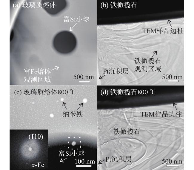
图 1 (a-b) 加热前,富铁熔体的HAADF图像和铁橄榄石的BF图像;(c-d)800 ℃时,富铁熔体的HAADF图像和np-Fe0颗粒的FFT图像,及铁橄榄石的BF图像Fig.1 (a-b) The HAADF of the Fe-rich melt and the BF images of fayalite before heating;(c-d) The HAADF image of the Fe-rich melt and the FFT pattern of np-Fe0 particle, as well as the BF image of fayalite at 800℃ |
2.2 辉石
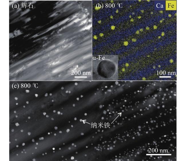
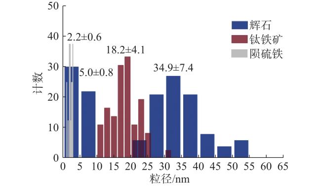
图 2 (a)加热前辉石的HAADF图像;(b-c) 800 ℃时,辉石的能谱面扫和纳米铁的HRTEM图像;以及持续恒温60分钟后的纳米铁粒径分布的HAADF图像Fig.2 (a) The HAADF image of pyroxene before heating; (b-c) The EDS mapping of pyroxene and the HRTEM image of np-Fe0 particle at 800 ℃, and the HAADF image of the size distribution of np-Fe0 particles at 800 ℃ for 60 min |
2.3 陨硫铁
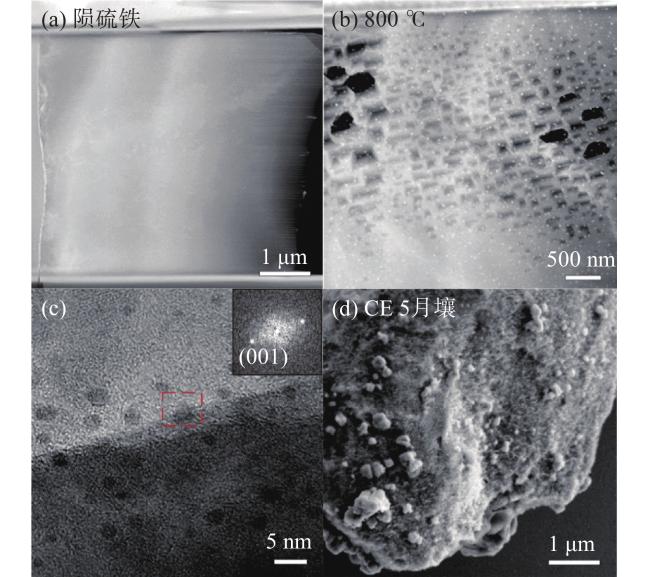
图 3 (a)陨硫铁加热前的HAADF图像;(b)800 ℃时,陨硫铁出现了六边形气孔和纳米铁;(c)陨硫铁的HRTEM图像和纳米铁的FFT图像;(d)嫦娥五号月壤中陨硫铁的表征结构Fig.3 (a) The HAADF image of troilite before heating;(b) There are hexagonal pores and the np-Fe0 in troilite at 800 ℃; (c) The HRTEM image of troilite and the FFT pattern of the np-Fe0; (d) Characterization and structure of troilite in the Chang'E-5 lunar soil |
2.4 钛铁矿
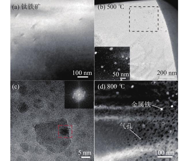
图 4 (a)钛铁矿加热前的HAADF图像;(b)500 ℃时, 钛铁矿的BF图像和纳米铁的HAADF图像;(c)纳米铁的HRTEM图像和FFT图像;(d)800 ℃时,钛铁矿中出现了气孔结构。Fig.4 (a) The HAADF image of ilmenite before heating; (b) The BF image of ilmenite at 500 ℃ and the HAADF image of np-Fe0 particles;(c) The HRTEM image and the FFT pattern of np-Fe0 particles; (d) There are pore structures in the ilmenite at 800 ℃ |